Rodents of multiple species in Europe, notably the bank vole (Myodes glareolus), common vole (Microtus arvalis), field vole (Microtus agrestis), yellow-necked field mouse (Apodemus flavicollis), Black Sea field mouse (Apodemus ponticus) and striped field mouse (Apodemus agrarius), harbour genetically distinct hantaviruses (genus Hantavirus, family Bunyaviridae) [Reference Vapalahti1–Reference Pounder3]. Some of these hantaviruses, such as Puumala virus and Dobrava virus, cause haemorrhagic fever with renal syndrome (HFRS) in Europe, while others, such as Tula virus and Tatenale virus, are not known to be pathogenic.
As evidenced by the detection of HFRS antigens by enzyme immunoassay, shrews and moles (order Soricomorpha, family Soricidae and Talpidae), captured in European Russia, were suspected of serving as reservoir hosts of hantaviruses more than 30 years ago [Reference Tkachenko4, Reference Gavrilovskaya5]. Although the specificity of such antigen-detection methods is uncertain, recent studies, using sensitive molecular-based technology, have unequivocally demonstrated that the hantaviruses hosted by several soricomorph species in Europe are far more genetically diverse than rodent-borne hantaviruses [Reference Song6–Reference Radosa11].
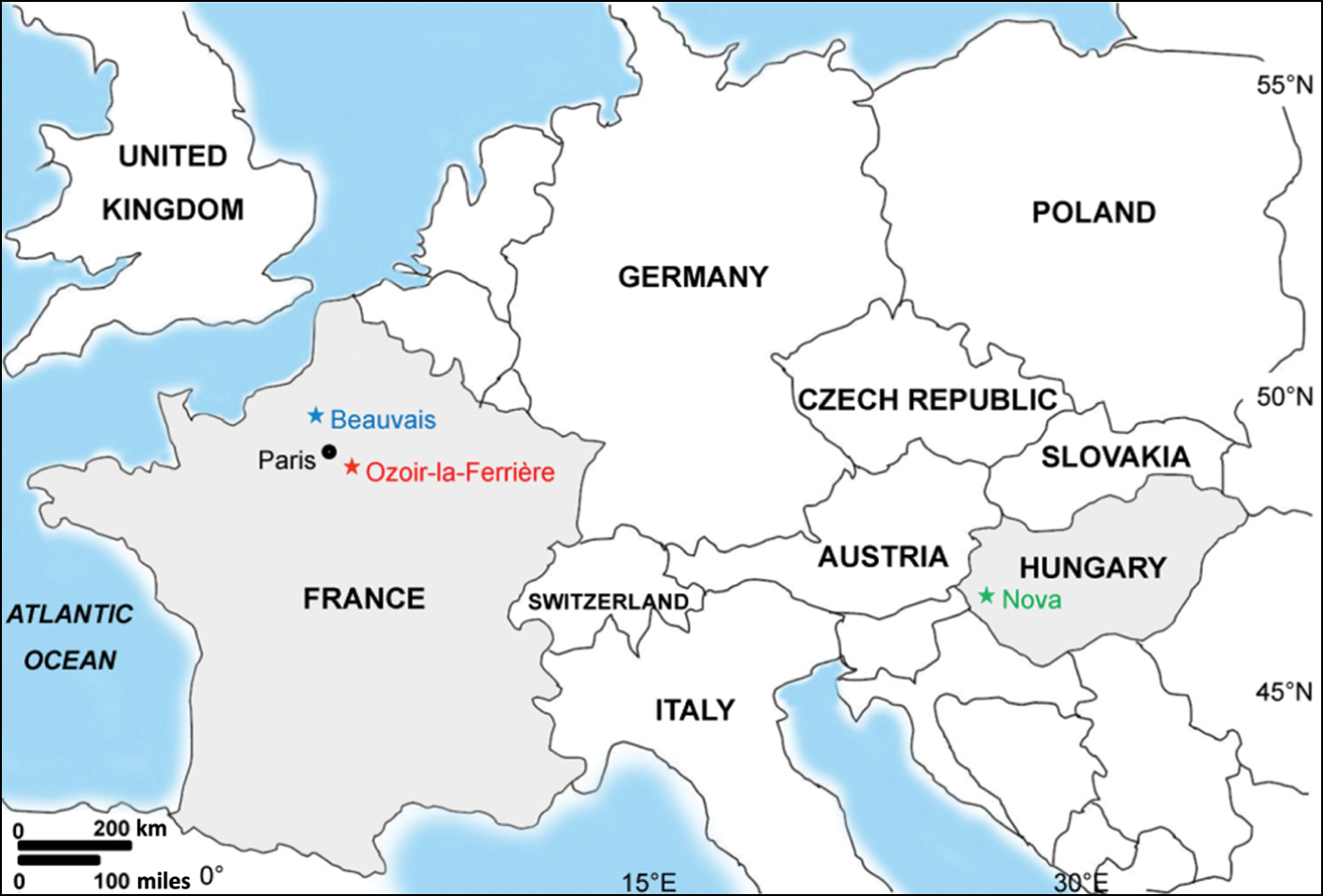
To date, one of the most highly divergent lineages of hantaviruses is represented by Nova virus (NVAV), detected in archival liver tissue from a single European mole (Talpa europaea), captured in Zala county, Hungary, in July 1999 [Reference Kang7]. To determine if this mole species is the true reservoir host of NVAV, European moles were captured using Putange mole traps [Reference Dormion12] during October 2012 to March 2013 in two venues frequented by humans: an 18-hole public golf course in Ozoir-la-Ferrière (48·7645°N, 2·6512°E), located 25·6 km east-southeast from metropolitan Paris in the Seine-et-Marne department of the Île-de-France region, and a recreational centre and nature park, called ‘Le Plan d'eau du Canada’, in Beauvais (49·4526°N, 2·0582°E), located in the Oise department in the northern French region of Picardy, ∼79 km from Paris (Fig. 1). All moles were frozen at −20°C for several days to months before processing. For tissue dissection, moles were partially thawed and lung tissues were removed using ethanol-cleaned instruments and placed in RNAlater® RNA Stabilization Reagent (Qiagen Inc., USA).

Fig. 1. Map of Europe, showing the locations of Ozoir-la-Ferrière (red) and Beauvais (blue) in France and Nova (green) in Hungary, where Nova hantavirus-infected European moles were trapped.
Total RNA was extracted, using the PureLink Micro-to-Midi total RNA purification kit (Invitrogen, USA), from lung tissues of 94 moles, and cDNA was prepared using the SuperScript III First-Strand Synthesis System (Invitrogen) and random hexamers. PCR was performed as described previously, using oligonucleotide primers designed from NVAV and other soricomorph-borne hantaviruses [Reference Song6, Reference Kang7, Reference Gu10], and DNA sequencing was performed using an ABI Prism 377XL Genetic Analyzer (Applied Biosystems, USA).
Using ClustalW, pairwise alignment and comparison of partial L-segment sequences of 362, 418 or 801 nucleotides (nt) revealed that 61 (64·9%) of the 94 moles were infected with NVAV. Of 36 and 28 moles captured in Ozoir-la-Ferrière on 18 October 2012 and 21 February 2013, 22 (61·1%) and 18 (64·3%), respectively, were NVAV positive, while 21 (70·0%) of 30 moles captured in Beauvais on 3 March 2013 were positive. In rodents, hantavirus infection is typically more prevalent in the heaviest males [Reference Vapalahti1, Reference Jonsson, Figueiredo and Vapalahti2]. By contrast, in European moles, NVAV was detected in 26/41 males (63·4%) and 35/53 females (66·0%), indicating no gender difference. Moreover, no statistically significant difference was found in infection rates in different weight classes.
Analysis of the full-length S segment of 11 NVAV strains (four from Ozoir-la-Ferrière, seven from Beauvais) revealed a single open reading frame, encoding a 428-amino acid nucleocapsid (N) protein (nucleotide positions 53–1336 nt), and 3′ and 5′ non-coding regions of 52 and 488 or 474 nt, respectively. The N protein secondary structures of the newly found NVAV strains were identical to that of prototype NVAV (data not shown) [Reference Kang7].
Although the partial L and full-length S segments of the NVAV strains from France differed from the prototype NVAV from Hungary by 13·6–16·5% and 12·1–14·2%, respectively, at the nucleotide level, they represented variants of the same virus, as evidenced by amino-acid sequence similarities of 96·6–98·1% and 97·0–97·4%, respectively (Table 1). Like the prototype NVAV, the NVAV strains from France diverged by ∼35% from all other rodent-, shrew- and mole-borne hantaviruses.
Table 1. Nucleotide and amino acid L- and S-segment sequence similarity (%) between newly identified NVAV strains from France and prototype NVAV from Hungary

nt, Nucleotide; aa, amino acid.
Sequence similarities of the 801-nucleotide partial L segment for only 11 of 61 newly found NVAV strains are shown.
The genetic diversity of the partial L segment in NVAV strains from Beauvais (0–13·1%) was much higher than that of NVAV strains from Ozoir-la-Ferrière (0–0·6%). The reasons for this are not entirely clear. One possibility is that because the trap site in Ozoir-la-Ferrière is a golf course, it might have undergone much more extensive habitat rearrangement and later become repopulated with a founder population of NVAV-infected European moles. By contrast, Beauvais represents a more natural and unaltered habitat with possibly greater diversity of European mole host genetics. This latter hypothesis is now being investigated.
Phylogenetic analyses based on partial L-segment sequences, using maximum-likelihood and Bayesian methods [Reference Kang7], showed that NVAV from France formed a highly divergent clade with prototype NVAV from Hungary (Fig. 2). A similar tree topology, well supported by bootstrap analysis and posterior node probabilities, was generated from analysis of the full-length S-segment sequences (Fig. 2). NVAV strains segregated along geographically specific lineages (Fig. 2), as reported previously for hantaviruses hosted by rodents and shrews.

Fig. 2. Phylogenetic trees generated by the maximum-likelihood and Bayesian methods, using the GTR+I+Γ model of evolution, based on the full-length S segment (S) and 801-nucleotide partial L segment (L) of Nova virus (NVAV). Since tree topologies were very similar using RAxML and MrBayes, the trees generated by MrBayes were displayed. The phylogenetic positions of prototype NVAV MSB95703 (green) (GenBank, S: FJ539168; L: FJ593498) from Hungary and the newly identified NVAV strains from Ozoir-la-Ferrière (red) (S: KF010573–KF010576; L: KF010535–KF010563) and Beauvais (blue) (S: KF010565–KF010571; L: KF010517–KF010534) in France are shown in relationship to representative hantaviruses harboured by crocidurine shrews, including Thottapalayam virus (TPMV VRC66412; S: AY526097; L: EU001330), Imjin virus (MJNV Cl05-11; S: EF641804; L: EF641806) and Jeju virus (JJUV 10-11; S: HQ834695; L: HQ834697). Also shown are soricine shrew-borne hantaviruses, including Cao Bang virus (CBNV CBN-3; S: EF543524; L: EF543525), Kenkeme virus (KKMV MSB148794; S: GQ306148), Seewis virus (SWSV mp70; S: EF636024), and Qiandao Lake virus (QDLV YN05-284; S: GU566021); and mole-borne hantaviruses, including Asama virus (ASAV N10; S: EU929072; L: EU929078) and Oxbow virus (OXBV Ng1453; S: FJ539166; L: FJ593497). Rodent-borne hantaviruses include Hantaan virus (HTNV 76-118; S: NC_005218; L: NC_005222), Soochong virus (SOOV SOO-1; S: AY675349; L: DQ056292), Dobrava virus (DOBV Greece; S: NC_005233; L: NC_005235), Seoul virus (SEOV 80-39; S: NC_005236; L: NC_005238), Tula virus (TULV M5302v; S: NC_005227; L: NC_005226), Puumala virus (PUUV Sotkamo; S: NC_005224; L: NC_005225), Prospect Hill virus (PHV PH-1; S: Z49098; L: EF646763), Sin Nombre virus (SNV NMH10; S: NC_005216; L: NC_005217) and Andes virus (ANDV Chile9717869; S: NC_003466; L: AF291704). The numbers at each node are posterior node probabilities based on 150 000 trees (left) and bootstrap values of 1000 replicates executed on the RAxML BlackBox web server (right), respectively. The scale bars indicate nucleotide substitutions per site.
Prevalence of anti-viral antibodies and/or viral antigens in rodent species known to harbour hantaviruses generally varies widely, depending on seasonal factors, reservoir population density, geographical location and ecological diversity [Reference Lee, Lee and Johnson13, Reference Dizney and Ruedas14]. The high prevalence of NVAV infection in European moles in France suggests efficient enzootic virus transmission and a well-established reservoir host–hantavirus relationship. Recently, a similarly high prevalence of NVAV infection, as evidenced by RT–PCR and confirmed by DNA sequencing, has been found in European moles captured in central Poland (S. H. Gu and R. Yanagihara, unpublished observations), strengthening the conjecture that this mole species serves as the reservoir of NVAV.
NVAV is probably widespread throughout the vast distribution of the European mole, which extends from the UK to Russia. Studies are needed to ascertain the influence of climate change, insect and predator abundance, and other factors on the dynamics and temporal fluctuations of NVAV transmission. Moreover, a foundation for disease forecasting may be warranted. That is, although neither NVAV nor any of the other recently discovered hantaviruses hosted by shrews and moles has been etiologically linked to diseases in humans, a similar situation existed two decades ago, before the realization that neotomine rodent-borne hantaviruses were capable of causing a rapidly progressive, frequently fatal disease, now known as hantavirus cardiopulmonary syndrome. As such, heightened awareness about NVAV would prompt physicians and public health workers to be on the alert for febrile illnesses or unusual clinical syndromes, occurring in mole catchers and field biologists, who are exposed to European moles and their secretions or excretions.
ACKNOWLEDGEMENTS
This research was supported in part by U.S. Public Health Service grants R01AI075057 from the National Institute of Allergy and Infectious Diseases and P20GM103516 (Centers of Biomedical Research Excellence) from the National Institute of General Medical Sciences, National Institutes of Health.
DECLARATION OF INTEREST
None.